Uterine Fibroids in Sub-Saharan Africa: Restoring Uterine Health and Fertility
Addressing the endemic 70-80% uterine fibroid burden among African women and the critical role of custom myomectomies and Vitamin D prevention.
The Endemic Burden of Leiomyomas
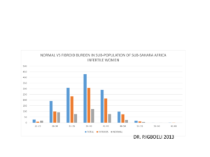
Uterine fibroids (leiomyomas) represent a significant medical burden, particularly for Black women who suffer from these benign tumors at much higher rates than other populations. In the poor-resource sub-Saharan region, it is estimated that 70% to 80% of women of child-bearing age develop uterine fibroids during their lifetime, leading to severe complications, primary or secondary infertility, and increased morbidity due to a lack of timely clinical care.
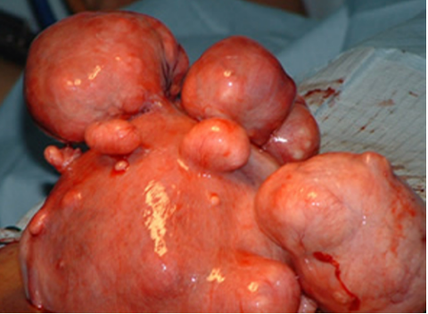
Uterine Distortion and Mechanical Infertility
Common symptoms include chronic pelvic pain, severe hemorrhage, and profound anemia that frequently requires multiple blood transfusions. When fibroids obstruct the uterine cavity or distort the fallopian tubes, they block natural paths of conception and increase miscarriage risks. The absence of properly trained surgical providers often leads to poor outcomes or unnecessary hysterectomies, cutting off a woman’s fertility prematurely.
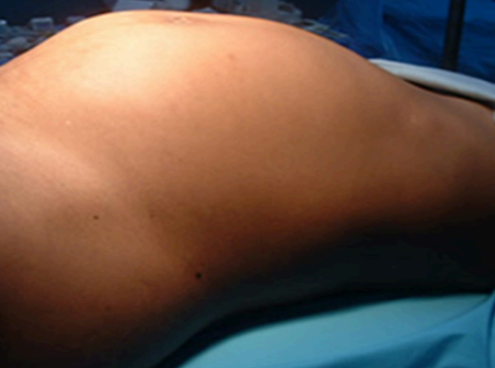
Precision Myomectomy and Reconstruction
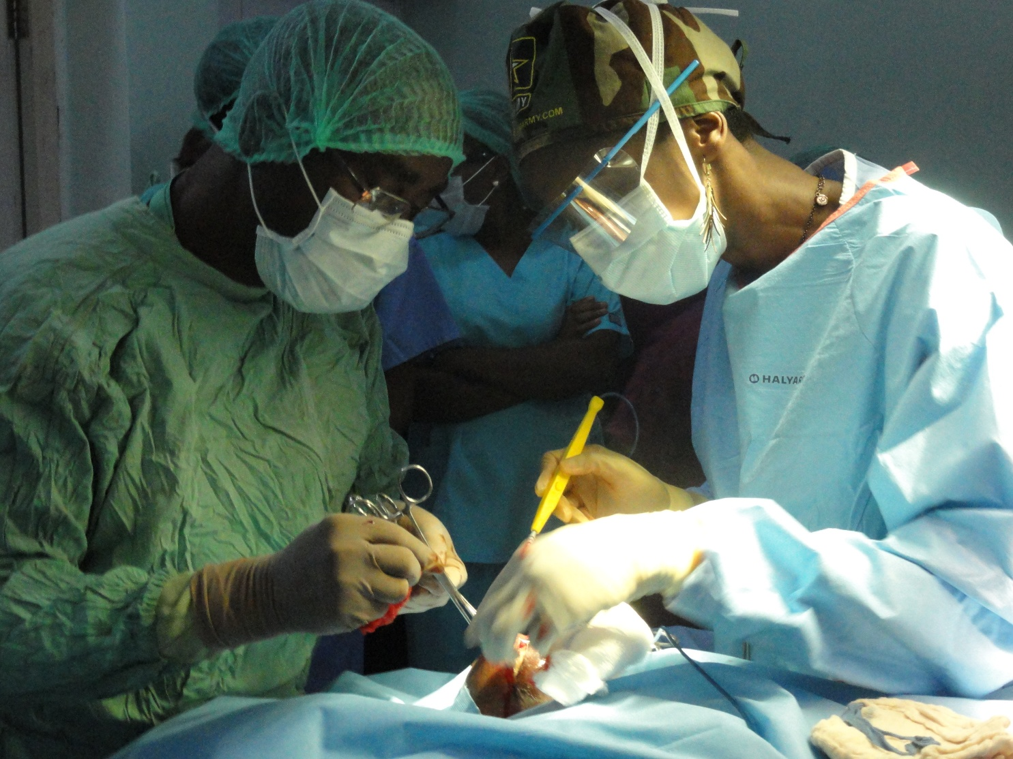
For women already presenting with large, symptomatic fibroids, advanced myomectomy (surgical removal) is the primary method to restore the womb’s structure. Prof. Prosper Igboeli and Dr. Ifeoma Igboeli FACS utilize micro-suturing techniques to reconstruct the uterine wall layer by layer, preserving reproductive function. This approach has allowed hundreds of patients to conceive naturally post-surgery.
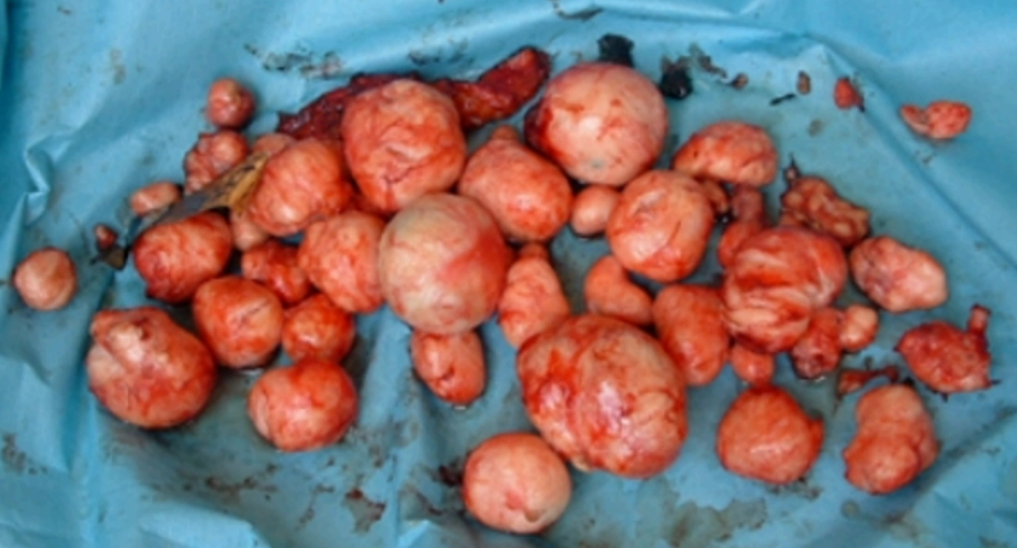
Uterine Restoration Case Success
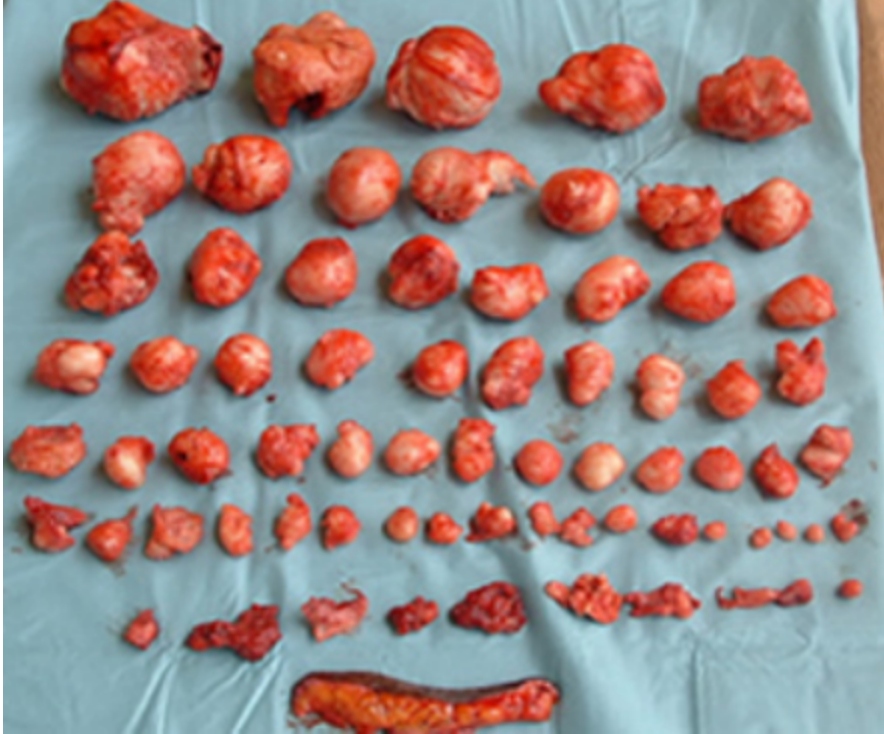
To ensure complete clearance and prevent recurrence, all enucleated fibroids are meticulously mapped and cataloged. This patient had over 60 fibroids successfully removed from her uterus. The uterine walls were reconstructed, restoring normal cavity architecture. Following recovery, patients under our protocol show excellent pregnancy rates and natural deliveries.
🧠 Key Takeaways
- ✅ Prevalence rates of 70-80% in women of African descent.
- ✅ Role of Vitamin D deficiency as a preventable risk factor.
- ✅ Restoration of pelvic cavity anatomy via specialized myomectomy.
- ✅ Uterine wall reconstruction preserving future gestation.